Gastroenterology
LEARN MORE
3D Colonoscopy & Gastroscopy
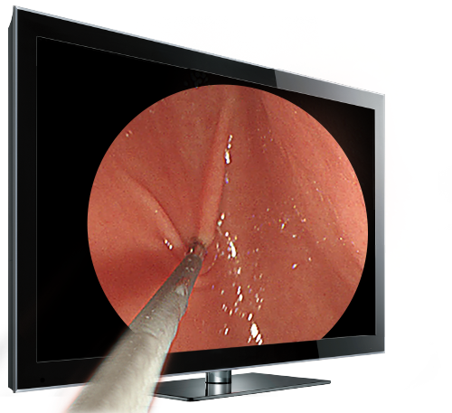
When it comes to identifying precancerous polyps, particularly non-polypoid and nonexcavated Type 0-II lesions, traditional flat 2D displays pose a challenge. However, prospective trial comparing the adenoma detection rates of 3D and 2D endoscopy within the team at National Taiwan University Hospital has revealed that 3D effectively increases the overall adenoma detection rate by 80% and elevates the detection rate of non-polypoid adenomas by 96%. This is due to the enhanced depth perception provided by stereoscopic vision, which makes it easier to spot adenomatous polyps, even those with slight elevation.
3D EMR
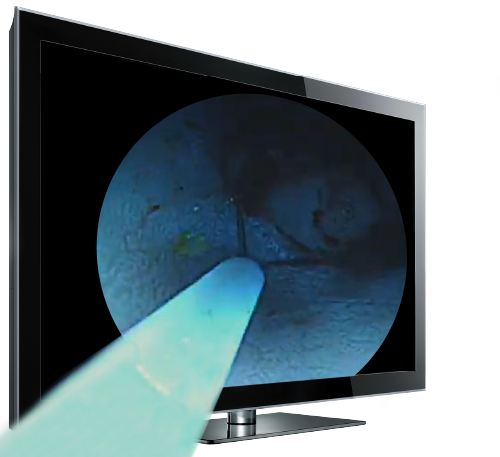
When utilizing an electrical snare for lesion removal, the integration of DARWIN 3D Visualization System significantly enhances procedural precision and efficiency. This advanced visual aid empowers medical professionals to meticulously resect abnormal tissue while preserving the integrity of surrounding healthy tissue. Within clinical environments, gastroenterologists have attested to the invaluable role of stereoscopic views in optimizing hand-eye coordination and distinguishing different tissue layers.
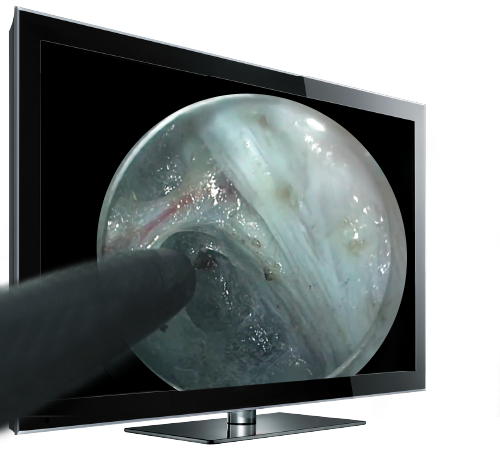
3D ESD
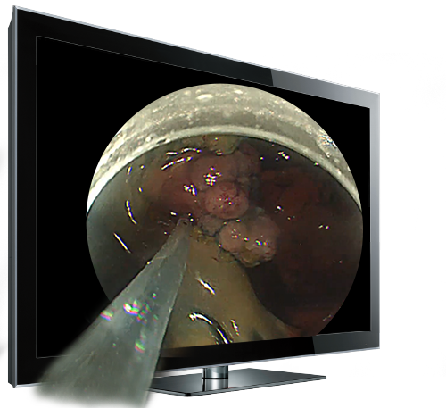
Performing procedures such as marking, dissection, and resection in the digestive system with occasional contraction demands precise techniques. Thanks to DARWIN 3D Visualization System, physicians can now conduct lesion assessments with greater ease and accuracy, thus lowering the chances of incomplete removal or perforation. DARWIN's stereoscopic view aids in simplifying tissue removal by providing clear spatial relationships, thereby minimizing the risk of unintended injury.
3D ERCP
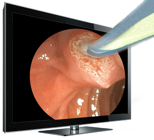
With DARWIN 3D Visualization System, gastroenterologists can navigate through tortuous or narrow ducts with depth perception, reducing the risk of complications such as perforation or duct injury. During cannulation, the stereoscopic view also widens the visual field, significantly aiding gastroenterologists in completing such a delicate procedure.
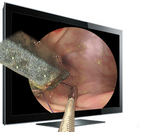
3D ESG
DARWIN 3D Visualization System enables doctors to achieve precise suture placement by providing clear visualization of depth and orientation relative to the gastric wall. This ensures optimal tissue apposition, mitigating the risk of complications like slippage or erosion. In fact, gastroenterologists acclaim DARWIN for its ability to elevate image accuracy and sharpness, contributing to greater surgical success rates.
3D POEM
3D visualization enhances depth perception and spatial awareness of gastroenterologists, facilitating smoother navigation through the esophageal layers during POEM. The DARWIN 3D Visualization System empowers doctors to manipulate instruments with heightened precision and control, especially during intricate tasks like creating the submucosal tunnel and conducting the myotomy. Furthermore, the enhanced spatial awareness leads to more efficient performance, ultimately decreasing procedural time and minimizing patient discomfort.